JACC: CardioOncology Editor-in-Chief Bonnie Ky is joined by Associate Editor Dr. Ron Witteles, Dr. Wilson Gonsalves, and Dr. Rola Khedraki in this insightful, informative two-part discussion on the topic of Amyloidosis. One of the Journal's most popular manuscripts to date, "AL Amyloidosis for the Cardiologist and Oncologist" is reviewed, with additional insights from recently published primers and clinical case challenges.
Explore papers referenced in Virtual Case Presentation Series on JACC.org: https://bit.ly/2AE18FI
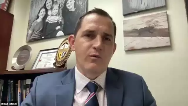
Joshua D. Mitchell, MD from Washington University in St. Louis speaks about Cardiovascular Manifestations From Therapeutic Radiation: A Multidisciplinary Expert Consensus Statement From the International Cardio-Oncology Society.
Link to Abstract-
https://www.jacc.org/doi/10.1016/j.jaccao.2021.06.003
Abstract
Radiation therapy is a cornerstone of cancer treatment, with more than half of patients receiving it. As a result of increased use and survival, researchers are paying more attention to the long-term effects of ionizing radiation, including cardiovascular damage. Radiation therapy can cause vascular atherosclerosis, as well as valve, cardiac, and pericardial dysfunction. The International Cardio-Oncology Society has issued a consensus statement based on broad principles of radiation treatment, cardiovascular risk assessment, and risk mitigation in this population. Anatomically based suggestions for cardiovascular management and follow-up are offered, with a focus on the early detection of atherosclerotic vascular disease on imaging to aid in the development of preventative medication. Radiation-induced cardiovascular disease has its own set of management issues, which are discussed as well. The diverse expert panel's recommendations are based on the most recent literature and constitute a unanimous consensus.
Highlights
• Radiation therapy causes valvular, myocardial, and pericardial illness, as well as other short- and long-term cardiovascular side effects in the vasculature and heart.
• Computed tomography scans used for radiation planning or cancer staging can be used to diagnose asymptomatic atherosclerosis and target preventive treatments.
• Additional practical cardiovascular disease screening suggestions based on anatomical exposure are offered.
• Radiation-induced cardiovascular disease requires special considerations, and modern percutaneous therapy is frequently favored over surgical methods.
Introduction
Since its first use in 1899, therapeutic radiation has become a cornerstone of cancer treatment, with more than half of cancer patients now receiving it (RT). RT has both short- and long-term side effects, despite its good impact on cancer outcomes and survival. RT has been related to an increased risk of long-term unfavorable cardiovascular (CV) consequences, which can lead to higher morbidity and death in cancer survivors, according to research (1-3).
Radiation-induced CV disease has a wide range of symptoms. Vascular illness (including coronary artery disease [CAD] and subclavian artery stenosis), valvular disease, constrictive pericarditis, restrictive cardiomyopathy, and heart failure (HF) are all considerably enhanced after thoracic RT. (Central Illustration). Patients are also at a greater risk of carotid artery stenosis and cerebrovascular accidents after head and neck or whole brain RT. Aorto-iliac atherosclerosis and renal artery stenosis are possible side effects of abdominal or pelvic RT. Deoxyribonucleic acid damage, oxidative stress, and the release of inflammatory and profibrotic cytokines all play a role in radiation-induced CV illness, leading to vascular, myocardial, valvular, and pericardial fibrosis.
In light of the entire context of RT benefits and hazards, doctors must be aware of CV problems and implement appropriate screening, mitigation, and prevention techniques into their practice. Although there are guidelines and expert consensus statements for detecting RT-related heart damage and dysfunction , there are few screening recommendations for extracardiac vascular symptoms. Furthermore, screening criteria for thoracic RT survivors have concentrated on the detection of obstructive CAD rather than nonobstructive CAD. This narrow focus overlooks the risk of myocardial infarction in patients with nonobstructive CAD and the importance of early medical intervention in these patients . Furthermore, newer imaging modalities such as coronary artery calcium (CAC) screening and coronary computed tomography (CT) angiography, which can more thoroughly identify a patient's CV risk during and after RT, are not included in current guidelines .
The current International Cardio-Oncology Society consensus statement systematically reviews available data and provides comprehensive recommendations for preventing, screening, diagnosing, and managing CV disease in cancer survivors receiving or who have received RT to the head and neck, chest, abdomen, and pelvis. This document discusses fundamental concepts for preventing CV illness following radiation therapy and offers recommendations for detecting specific vascular effects based on anatomical location.
The International Cardio-Oncology Society was founded in October 2019 as an international, multidisciplinary collaboration of professionals in medical oncology, radiation oncology, cardiovascular imaging, and cardio-oncology. A comprehensive literature analysis was conducted by searching the PubMed index for studies published in English from 1975 to the present, combining text words and Medical Subject Headings such as radiation, cardiotoxicity, and CV anomalies; the final search yielded 2,999 publications. The literature evaluation was completed in October 2019 and then revised before the recommendations were finalized in December 2020. We also looked for relevant titles in the references of the publications we reviewed. Evidence from randomized controlled trials or meta-analyses was given top priority, followed by well-designed nonrandomized research, other nonrandomized studies, expert opinion, and clinical experience. ClinicalTrials.gov was also used to look for any relevant clinical trials.
To study the material and provide practical recommendations, the committee met bimonthly via webinars and teleconferences. Recommendations are tagged with language like "is advised," "may be useful," and "can be considered," according to American College of Cardiology/American Heart Association consensus statements (18). Because there are few randomized controlled trials particularly designed to address CV disease in RT survivors, no levels of evidence are provided. These proposals are the result of a consensus among the members of the multidisciplinary expert committee.
Stefan Janssens, MD-
Link to Article- https://www.jacc.org/doi/10.1016/j.jacbts.2021.07.001
• It is uncertain what role CLEC4E plays in myocardial repair following ischemia-reperfusion damage.
• Deletion of CLEC4E is linked to decreased cardiac damage, inflammation, and structural and functional remodeling of the left ventricle.
• CLEC4E is a promising target for reducing myocardial inflammation and improving ischemia-reperfusion damage repair.
Summary
C-type lectin domain family 4 member E (CLEC4E) from bacteria has a vital function in sterile inflammation, although its significance in myocardial healing is uncertain. We show that CLEC4E expression levels in the myocardium and blood correspond with the extent of myocardial injury and left ventricular (LV) functional impairment using complementary techniques in porcine, murine, and human tissues. In the ischemic heart, CLEC4E expression is dramatically enhanced in the vasculature, cardiac myocytes, and infiltrating leukocytes. A reduction in acute cardiac damage, neutrophil infiltration, and infarct size has been linked to the loss of Clec4e signaling. At 4 weeks, Clec4e–/– had considerably enhanced LV structural and functional remodeling due to reduced myocardial damage. The early transcriptome of Clec4e–/– mice LV tissue revealed considerable overexpression of transcripts involved in myocardial metabolism, radical scavenging, angiogenesis, and extracellular matrix organization compared to wild-type mice. As a result, targeting CLEC4E in the early stages of ischemia-reperfusion injury offers a prospective therapeutic method for modulating myocardial inflammation and enhancing myocardial healing.
Introduction
After a myocardial infarction, cardiac repair is a highly organized and complex series of processes that begins with an inflammatory phase marked by immune cell infiltration and the production of danger-associated molecular patterns (DAMPs) and then progresses to a reparative and proliferative phase (1-3). To allow later tissue healing and prevent maladaptive left ventricular (LV) remodeling, faulty scar formation, and poor patient outcome, the early inflammatory response with DAMPS binding to cognate pattern recognition receptors (PRRs) must be correctly and timely coordinated (4,5). Excessive or protracted inflammation causes ventricular dilatation and systolic dysfunction, increasing the likelihood of heart failure (6), whereas insufficient early inflammation fails to remove necrotic cardiac cells and matrix components, obstructing eventual tissue healing. The importance of a better understanding of the cellular and molecular pathways directing this biphasic repair process following ischemia-reperfusion (I/R) injury is highlighted by this complex and tightly regulated innate immune activation response.
Previously, we looked examined how the transcriptional profile of patients with an acute myocardial infarction (AMI) changed over time in circulating blood cells (7). We found that pro-inflammatory pattern recognition receptors, such as the C-type lectin domain family 4 member E receptor (CLEC4E), which is normally expressed on leukocytes and activated in response to bacteria, were highly activated (8,9). In vitro, this innate immune receptor detects necrotic material caused by ischemia injury (10) and suppresses the inflammatory response in experimental murine brain injury (11,12). CLEC4E's role in myocardial I/R damage, on the other hand, is uncertain.
The significance of CLEC4e signaling in the early inflammatory response and subsequent healing phase following acute myocardial ischemia injury was studied in this study, as well as its potential as a new target for intervention in myocardial I/R injury.
Methods
Ethics
The Ethics Committee for Animal Experimentation at KU Leuven (P244/2014 and P064/2017) approved all animal procedures, which were carried out in accordance with Belgian law on the care and use of experimental animals. The patient research procedure followed the Declaration of Helsinki, and all patients signed informed permission (Ethical Committee ML8525, Belgian trial no. B322201214942, S54129) (7).
I/R damage in a porcine model and cardiac magnetic resonance imaging
As previously documented, ten domestic pigs (body weight: 20-30 kg) were sedated, anesthetized, and had I/R damage by transient balloon blockage of the left anterior descending coronary artery (LAD) distal to the first diagonal branch for 50 minutes, followed by 4 hours of reperfusion (7). After 4 hours of reperfusion, a subgroup of 6 pigs underwent 3-T cardiac magnetic resonance imaging (MRI) (Prisma-Tim, Siemens) to assess infarct size (MI/left ventricle), end-systolic volume (ESV), end-diastolic volume (EDV), and ejection fraction, before being euthanized (13). Biopsy specimens were taken from the left ventricle's ischemia, border, and distant zones for differential gene expression analysis and histology. Supplementary Materials and Methods, as well as Supplementary Table 1, give more information.
I/R damage in a mouse model and cardiac MRI
Male C57Bl6/J wild-type (WT) mice aged 12 to 14 weeks were crossed with Clec4e–/– mice (031936-UCD) purchased from the Mutant Mouse Resource and Research Center. Mice were given I/R damage by ligating the LAD for 60 minutes and then reperfusing them, as described before (14). They were randomly assigned to evaluate arms with reperfusion for 24 hours (WT, n = 12; Clec4e–/–, n = 14), 72 hours (WT, n = 5; Clec4e–/–, n = 5) or 4 weeks (WT, n = 17; Clec4e–/–, n = 16). Investigators blinded to the genotype of the animals obtained cardiac MRI data on a Bruker BioSpec 70/30 7T MRI machine (Bruker BioSpin) after 4 weeks of reperfusion. Blood was taken from the inferior caval vein after euthanasia to measure high-sensitivity troponin I (hs-TnI) as a surrogate marker of cardiac injury and to isolate neutrophils to study their phenotype and migration capacity; at 24 hours after I/R injury, LV tissue was taken for histological analysis and transcriptome studies. Organs were perfused for 10 minutes with saline before being harvested for histological and differential gene expression analyses utilizing ribonucleic acid (RNA) sequencing and quantitative real-time polymerase chain reaction (qRT-PCR). The Supplementary Materials and Methods section has more information.
June-Wha Rhee, MD, cardiologist with specialized clinical and research training in cardiovascular drug toxicity and pharmacogenomics, from Stanford Hospital and Clinics speaks about Impact of Hormonal Therapies for Treatment of Hormone-Dependent Cancers (Breast and Prostate) on the Cardiovascular System: Effects and Modifications.
Link to Abstract:
https://www.ahajournals.org/doi/pdf/10.1161/HCG.0000000000000082
Overview:
The main causes of mortality in the United States are cardiovascular disease and cancer, with hormone-dependent tumors (breast and prostate cancer) being the most frequent noncutaneous malignancies in women and men, respectively. The hormonal (endocrine-related) treatments that are used to treat both malignancies enhance survival rates, but they significantly raise cardiovascular morbidity and death in survivors. This consensus statement outlines the dangers of particular hormone treatments used to treat breast and prostate cancer, as well as an evidence-based strategy for preventing and detecting negative cardiovascular events. The cardiovascular effects of various durations of hormonal therapy, the cardiovascular risks associated with combinations of newer generations of more intensive hormonal treatments, and the specific cardiovascular risks that affect individuals of various races/ethnicities are all highlighted as areas of uncertainty.
Finally, a multidisciplinary approach to the application of lifestyle and pharmaceutical treatments for risk reduction and management is emphasized both during and after active therapy.
June-Wha Rhee, MD, a cardiologist with specialized clinical and research training in cardiovascular drug toxicity and pharmacogenomics, from Stanford Hospital and Clinics speaks about Impact of Hormonal Therapies for Treatment of Hormone-Dependent Cancers (Breast and Prostate) on the Cardiovascular System: Effects and Modifications.
Link to Abstract:
https://www.ahajournals.org/doi/pdf/10.1161/HCG.0000000000000082
Overview:
The main causes of mortality in the United States are cardiovascular disease and cancer, with hormone-dependent tumors (breast and prostate cancer) being the most frequent noncutaneous malignancies in women and men, respectively. The hormonal (endocrine-related) treatments that are used to treat both malignancies enhance survival rates, but they significantly raise cardiovascular morbidity and death in survivors. This consensus statement outlines the dangers of particular hormone treatments used to treat breast and prostate cancer, as well as an evidence-based strategy for preventing and detecting negative cardiovascular events. The cardiovascular effects of various durations of hormonal therapy, the cardiovascular risks associated with combinations of newer generations of more intensive hormonal treatments, and the specific cardiovascular risks that affect individuals of various races/ethnicities are all highlighted as areas of uncertainty.
Finally, a multidisciplinary approach to the application of lifestyle and pharmaceutical treatments for risk reduction and management is emphasized both during and after active therapy.
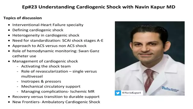
We discuss the various aspects of cardiogenic shock including definition, heterogeneity, classification and management with an internationally renowned expert in the field Dr Navin Kapur. Dr Kapur is the Director of the Acute Mechanical Circulatory Support Program at Tufts University School of Medicine, Boston, MA.
Listen, like, subscribe, and give us a high rating.
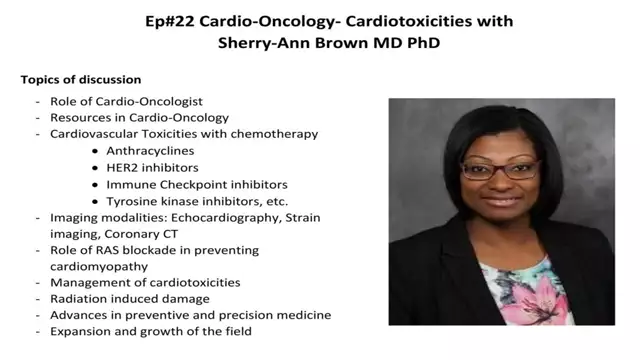
We discuss several aspects of cardio-oncology including screening protocols, specific chemotherapy related cardiotoxicities and management of cardiovascular disease among cancer patients and cancer survivors. Dr Sherry-Ann Brown is the director of Cardio-Oncology Medical College of Wisconsin, Milwaukee, WI.
Listen, like, subscribe, and give us a high rating.
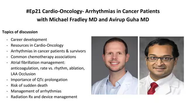
We discuss several aspects of cardiac arrhythmias and their management among cancer patients and cancer survivors with Drs Fradley and Guha. Dr Fradley is an electrophysiologist and Dr Guha is a non-invasive cardiologist, both with tremendous expertise in cardio-oncology. Dr Fradley is the Medical Director of Cardio-Oncology at University of Pennsylvania, Philadelphia, PA.
Listen, like, subscribe, and give us a high rating.
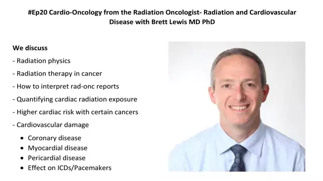
We discuss the physics of radiation, role for ionizing radiation in treating cancer & related cardiac damage with Dr Brett Lewis MD PhD. Dr. Lewis has interest & expertise in treating several thoracic malignancies & practices at Hackensack Meridian Health, NJ.
Listen, like, subscribe, and give us a high rating.
We discuss several classes of cancer therapies including chemotherapy, immunotherapy, CAR T-Cell therapies and related cardiovascular toxicity from the perspective of a board certified oncologist Dr Kaushal Parikh, MD. Dr Parikh specializes in thoracic oncology and practices at Hackensack Meridian Health, NJ.
Listen, Like, Subscribe, and give us a high rating!
Mehmet Asim Bilen, MD from Winship Cancer Institute of Emory University discusses the Evaluation of a novel blood pressure scoring method and its association with clinical response in cancer patients treated with anti-vascular endothelial growth factor therapy.
Link to Study -
https://pubmed.ncbi.nlm.nih.gov/24764123/
Instract
Background: In advanced cancer patients treated with anti-vascular endothelial growth factor (VEGF) therapies, the aim of this study was to develop a novel blood pressure (BP) scoring method and correlate it with clinical response.
Methods: We retrospectively evaluated data from 23 clinical trials involving at least one anti-VEGF agent for 368 patients. Using the standard Common Terminology Criteria for Adverse Events (CTCAE) version 4.0 and our latest system, BP scores were measured using both BP readings and the amount of anti-hypertensive drugs obtained by the patient. BP ratings at baseline and four months have been classified. Elevated scores were correlated with the clinical response through logistic regression analysis. An agreement was assessed between the CTCAE and the new system.
Results: Under the latest BP scoring system, partial response rates were 20 percent for four-month elevated patients versus 6 percent for non-elevated patients (P < 0.001). When adjusted for tumor type, age, sex, history of anti-VEGF treatment, and the number of anti-VEGF treatments, the odds ratio was 3.8 (95% confidence interval [CI]: 1.8, 8.2; P < 0.001) for elevated BP under the new scoring system. The kappa statistics were 0.57 (95 percent CI: 0.47, 0.67; P < 0.001) for agreement between the CTCAE and current scoring methods, suggesting substantial concordance.
Conclusion: The rise in BP scores was a proxy for favorable clinical response in patients receiving anti-VEGF therapy using the novel scoring system. Ultimately, this new approach offers details about the clinical tumor response over and above that given by the scoring method of CTCAE.
Journal Editor-in-Chief Bonnie Ky, MD, MSCE, FACC, moderates a video case presentation on Early Immune Checkpoint Inhibitor Cardiotoxicity presented by Han Zhu, MD, with associate editor discussants Dan Lenihan, MD, FACC; Ana Barac, MD, PhD, FACC; Ronald Witteles, MD, FACC; and Anne Blaes, MD. The experts discuss two newly published manuscripts related to the topic, “The Evolving Immunotherapy Landscape and the Epidemiology, Diagnosis, and Management of Cardiotoxicity,” by Lili Zhang, MD, ScM and Tomas G. Neilan, MD, MPH et al., and the French Working Group’s Viewpoint on “Management of Immune Checkpoint Inhibitor-Induced Myocarditis: Plea for a Pragmatic Approach,” led by Franck Thuny, MD, PhD, et al. Please visit https://www.jacc.org/ and https://www.acc.org/ for more information